PBMC - Peripheral Blood Mononuclear Cell
Eine periphere mononukleäre Blutzelle (Peripheral Blood Mononuclear Cell, PBMC) ist eine Blutzelle mit einem einzigen, runden Kern. Der Begriff „peripher“ bezeichnet ausgereifte Blutzellen, die sich nicht in blutbildenden Organen wie dem Knochenmark befinden, sondern im peripheren Blut zirkulieren.
NOTE: Unter peripherem Blut versteht man das Blut, das nach seiner Bildung im Knochenmark in die Blutgefäße gelangt und dort im Kreislauf zirkuliert.
Zu den Hauptbestandteilen des peripheren Blutes gehören neben dem Blutplasma, das als Transportmedium dient, auch rote Blutkörperchen (Erythrozyten), weiße Blutkörperchen (Leukozyten) ) und Blutplättchen (Thrombozyten).
PBMCs, sometimes referred to as PMNCs (peripheral mononuclear cells) or MNCs (mononuclear cells), comprise all mononuclear leukocytes. These include:
- Lymphocytes (T-cells, B-cells, NK-cells)
- Monocytes
- Dendritic Cells and
- Basophilic Granulocytes.
Isolation / enrichment of PBMC by density gradient centrifugation
The term PBMC (peripheral blood mononuclear cells) is closely associated with the isolation of cells from whole blood through a Density gradient centrifugation. Die Trennung der Blutbestandteile ist möglich, da sie verschiedene Dichten aufweisen; dies geschieht durch eine einstufige Zentrifugation in einem geeigneten Dichtegradientenmedium. Die Trenngrenze (Cut-Off) für die mononukleären Zellen liegt bei 1,077 g/ml.
Die dichtegradienten-angereicherten PBMC werden manchmal als Buffy Coat bezeichnet. In diesem Zusammenhang beschreibt Buffy Coat die Schicht der angereicherten Leukozyten in der Interphase. Für diesen speziellen Fall besteht der Buffy Coat nur aus mononukleären Zellen und ist in diesem Sinn ein spezieller Buffy Coat.
Polymorphonuclear leukocytes (with multiple segmented nuclei), such as granulocytes, are depleted in this fraction. Neutrophil and eosinophil granulocytes cannot be enriched from a PBMC fraction prepared by standard density gradient separation (the medium has a density of 1.077 g/ml). Basophilic granulocytes differ in density and can be partially found in the PBMC fraction. Erythrocytes are strongly reduced compared to the initial cell count.
First, a synthetic polymer solution with a defined density is overlaid with blood or sample material. After preparation, a centrifugation step takes place. In the interphase between the two solutions, the cells accumulate at the corresponding density. The interphase containing the PBMCs is carefully removed. The enriched mononuclear cells are then washed and used for further application.
Allgemeiner Ablauf einer PBMC Anreicherung mit Hilfe eines Dichtegradientenmediums
Zunächst wird eine synthetische Polymerlösung, mit einer definierten Dichte, mit Blut oder Probenmaterial überlagert. Nach der Vorbereitung erfolgt ein Zentrifugationsschritt. In der Interphase zwischen den beiden Lösungen reichern sich die Zellen mit der entsprechenden Dichte an. Die Interphase, die die PBMCs enthält, wird vorsichtig entfernt. Die angereicherten mononukleären Zellen werden anschließend gewaschen und für die weitere Anwendung verwendet.
Isolation / enrichment of PBMC with PBMC Spin Medium
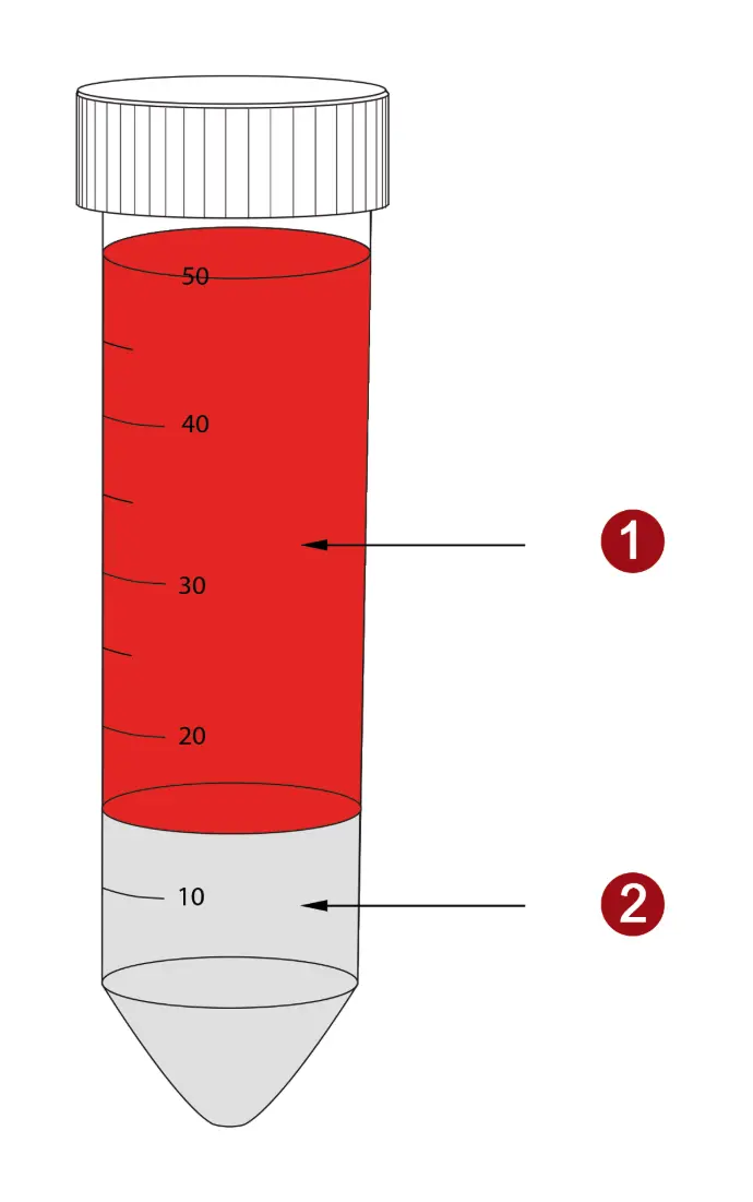
The whole blood is carefully overlayed to the PBMC Spin Medium (2), which is placed in a centrifuge tube. Care must be taken to ensure that the two liquids do not mix. Alternatively, pluriMate tubes can be used. The pluriMate barrier facilitates layering and prevents the sample material from mixing with the density medium.

Fig. 1: PBMC preparation with PBMC Spin Medium. The sample material (1) is carefully overlayed on the density medium (2).
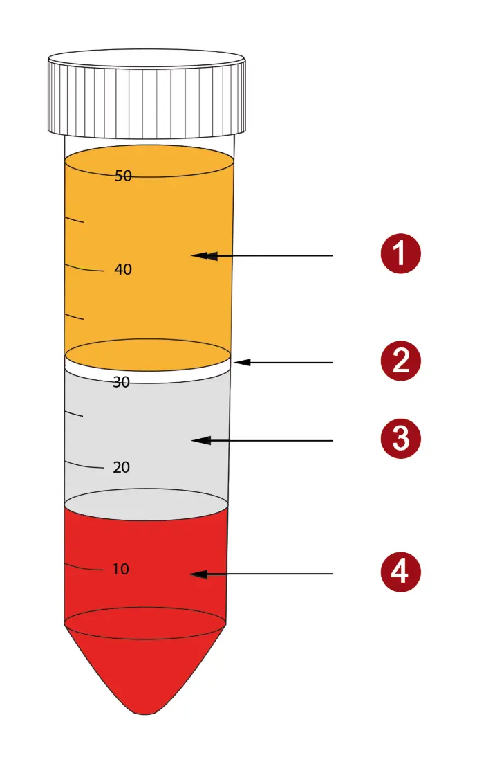
In the following centrifugation step, the actual density gradient centrifugationthe sample is separated into the different layers. The mononuclear cells (2) are enriched between the plasma (1) and the density medium (3).
